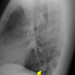
X-RAY CHEST LATERAL
Tested By Sri Divya Diagnostic Center
In a chest X-ray, an X-ray machine sends a beam of radiation through the chest, and an image is recorded on special film or a computer. This image includes organs and structures such as the heart, lungs, large blood vessels, diaphragm, part of the airway, the upper spine, ribs, collarbone, and breastbone. Usually, the X-ray technician will take pictures of the chest: • from the back of the chest (if the child is old enough to stand up for the X-ray) • from the side For younger children, th