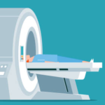
MRI CHEST
Tested By Sri Divya Diagnostic Center
What is MRI of the Chest? Magnetic resonance imaging (MRI) is a noninvasive test doctors use to diagnose medical conditions. MRI uses a powerful magnetic field, radiofrequency pulses, and a computer to produce detailed pictures of internal body structures. MRI does not use radiation (x-rays). Detailed MR images allow doctors to examine the body and detect disease. MRI of the chest gives detailed pictures of structures within the chest cavity, including the mediastinum, chest wall, pl
Lab Test Details
Description
Why is MRI- Chest done?
To diagnose myocardial perfusion (blood flow pattern to the heart) and myocardial infarction (scarring of the heart muscles due to poor flow or blockage)
To detect lymphatic, vascular (blood vessels) and pericardial disease (heart sac arond heart)
To find out tumor/cancer size of the heart (valves), lungs, and blood flow pattern of in the blood vessels of the heart and its chamber
To evaluate chest bones fracture/damage (ribs, collar bone, vertebrae) and its surrounding soft tissues, lesion of the chest.
How the Test is Performed ?
The test is done in the following way:
You may be asked to wear a hospital gown or clothing without metal fasteners (such as sweatpants and a t-shirt). Certain types of metal can cause blurry images or be dangerous to have on in the scanner room.
You lie on a narrow table, which slides into the large tunnel-shaped scanner.
You must be still during the exam, because movement causes blurred images. You may be told to hold your breath for short periods.
Some exams require a special dye called contrast. The dye is usually given before the test through a vein (IV) in your hand or forearm. The dye helps the radiologist see certain areas more clearly. A blood test to measure your kidney function may be done before the test. This is to make sure your kidneys are healthy enough to filter the contrast.
During the MRI, the person who operates the machine will watch you from another room. The test most often lasts 30 to 60 minutes, but it may take longer.
How to Prepare for the Test
You may be asked not to eat or drink anything for 4 to 6 hours before the scan.
Tell your provider if you are claustrophobic (afraid of closed spaces). You may be given a medicine to help you feel sleepy and less anxious. Your provider may suggest an "open" MRI, in which the machine is not as close to your body.
Before the test, tell your health care provider if you have:
Brain aneurysm clips
Artificial heart valves
Heart defibrillator or pacemaker
Inner ear (cochlear) implants
Kidney disease or are on dialysis (you may not be able to receive contrast)
Recently placed artificial joints
Vascular stents
Worked with sheet metal in the past (you may need tests to check for metal pieces in your eyes)
The MRI contains strong magnets, so metal objects are not allowed into the room with the MRI scanner. This is because there is a risk that they will be drawn from your body toward the scanner. Examples of metal objects you will need to remove are:
Pens, pocket knives, and eyeglasses
Items such as jewelry, watches, credit cards, and hearing aids
Pins, hairpins, and metal zippers
Removable dental work
Some of the newer medical devices described above are MRI compatible, so the radiologist needs to check the device manufacturer to determine if an MRI is possible.
₹ 8000.00 ₹ 7600.00
-
Free Shipping
For orders from ₹500 -
-
-
