Your Location
Call us
7799111544
User Image

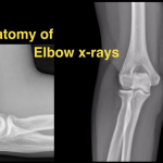
X-RAY ELBOW APLATERAL

Tested By Sri Divya Diagnostic Center

In an elbow X-ray, an X-ray machine sends a beam of radiation through the elbow, and an image is recorded on special X-ray film or a computer. This image shows soft tissues and bones of the elbow, including the humerus (the upper bone of the elbow joint), and the radius and ulna (the lower bones of the elbow joint). An X-ray technician will take pictures of the elbow: • from the front (anteroposterior, or AP, view) • from the side (lateral view) • at an angle (oblique view) Occasionall

Lab Test Details


Description

Why Are Elbow X-Rays Done?
An elbow X-ray can help doctors find the cause of pain, tenderness, swelling, or a deformity. It can also check for broken bones or a dislocated joint. After a broken bone has been set, an X-ray can help show if the bones are aligned and if they have healed properly.

An X-ray can help doctors plan surgery, when needed, and check the results after it. It also can help detect cysts, tumors, or other diseases in the bones, including later stages of bone infections.

₹ 400.00 ₹ 320.00

+
  • Sample Type scan
  • Free Shipping
    For orders from ₹500